Age Related Macular Degeneration

Vitrectomy

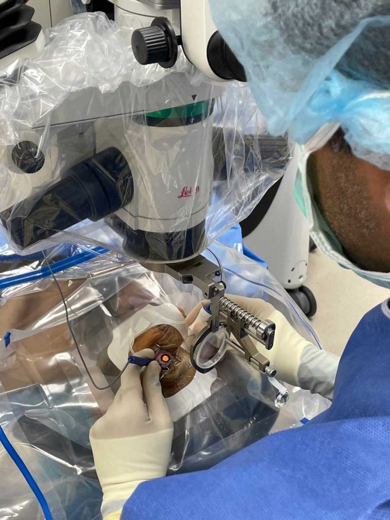
What is a Vitrectomy?

A vitrectomy is the surgical removal of the vitreous gel from the inside of the eye and is often done to repair or prevent traction retinal detachment, repair very large tears in the retina, reduce vision loss caused by blood in the vitreous, and to treat severe proliferative retinopathy that results in scar tissue formation or growth of new blood vessels on the retina despite attempts at laser treatment.

How is the procedure preformed?

During a vitrectomy the ophthalmologist inserts small instruments into the eye and cuts the vitreous gel and removes it. After removing the gel the physician can then more easily reach the retina and treat it with a laser, cut or remove fibrous scar tissue, flatten out areas where the retina may have detached, or repair tears or holes in the retina or macula. At the end of the retinal repair a gas bubble is injected into the eye to keep the retina pressed against the eye wall to reattach. Think of the retina as wallpaper that needs to be re-glued to the wall, the gas bubble serves to make sure the retinal layer stays in contact with the eye wall and reattaches seamlessly.

What to expect after surgery?

At Texoma Retina Center the Vitrectomy is preformed as an outpatient surgery. Often, following the surgery, the patient may have to position themselves so that the gas can push against the back of the eye to make sure the retina reattaches. Most likely the patient will be asked to keep their head facing down for a matter of hours per day. It is extremely important that patients listen and follow the post-operative instructions given to ensure proper healing. Because we ask to see our outpatient surgery patients the morning after their procedures, it may be easier for patients and families from out of town to stay in the Denison area, please visit our Hospitality and Extended Visits Page for information on hotels travel services.

Does it work and what are the risks?

Vitrectomy has been shown to greatly improve visual acuity in many people who have vitreal hemorrhages (blood in the vitreous). In general, surgery can restore some vision that is lost as a result of traction retinal detachments and can help prevent further detachments. The results tend to be better when the retinal detachment has not reached the macula, the area responsible for central, sharp vision. Each case is different, listening to the physician and asking knowledgeable questions provides the patient and family with the best information available

The risks associated with a vitrectomy include increased intraocular pressure, especially in patients with glaucoma. Other risks include further bleeding into the vitreous gel, retinal detachment, fluid build up in the cornea, infection inside the eye, and cataract formation. While risks are rare, they are always a possibility and something to think about.

For more questions about Vitrectomy procedures please visit WedMD’s article on Vitrectomy

Macular degeneration is currently treated by anti-VEGF drug injections into the eye every 6-12 months. These injections allow patients to keep their vision, but must be continually given. Research has found promising results using eye drops instead of injections, so that patients may soon have the convenience of administering their AMD treatment at home.

Request an
Appointment

At Texoma Retina and Glaucoma, our focus is on providing the most progressive cutting-edge vitreo-retinal services in a caring and compassionate atmosphere.

Opening Hours

Calls are taken 24/7 for emergencies

MONDAY
08.00 AM – 05.00 PM
TUESDAY
08.00 AM – 05.00 PM
WEDNESDAY
08.00 AM – 05.00 PM
THURSDAY
08.00 AM – 05.00 PM
FRIDAY
08.00 AM – 05.00 PM

Available on Saturday and Sunday for Emergency only. Call for appointment

LOCATIONS AND DIRECTIONS